Peri-Acetabular Osteotomy (PAO)
LOADING
A peri-acetabular osteotomy (PAO) is an extremely complex and technically difficult operation where an orthopaedic surgeon will carefully chisel out a portion of bone which contains the patients hip socket (“acetabulum”) from the rest of their pelvis, then adjust the position of the cut out acetabulum relative to that pelvis, and then finally pass pins from the pelvis into the re-directed portion of bone with the acetabulum to hold it in its new orientation and allow the cut pieces of bone to heal together.
There are two indications for this operation, the main one being a condition called “hip dysplasia”. This condition is characterised by the hip joint having an abnormal relationship between the proximal femur (the ball) and the pelvic acetabulum (the socket). There is a strong genetic linkage (i.e. it is often inherited), and is largely thought to occur during development of the foetus or early after childbirth. There are now neonatal and even foetal screening programmes in a large number of first world countries to detect this in foetuses and newborn babies, as treatment of this condition in the newborn can often prevent severe disability later in life. However, there are a proportion of the population who “slip through the net” of neonatal screening and only have their hip dysplasia detected much later in life, i.e. in their late 20’s up to early 40’s. These are usually patients who have much a more subtle form of the condition which makes it harder to detect when very young.
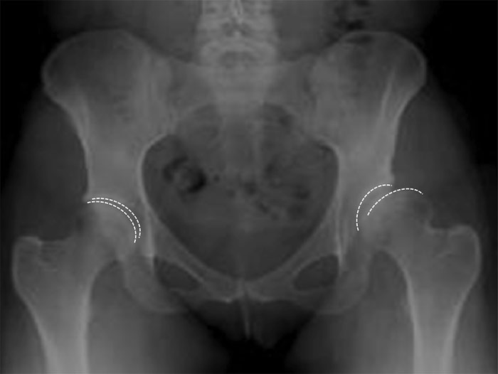
X-ray of Hip Dysplasia
The severity of this abnormal relationship is on a fairly wide spectrum. In severe cases there may be such gross abnormalities of the socket and the ball such that the socket is clearly mis-shapen and facing the wrong direction. In these cases, the acetabulum is shaped like a flat, upwardly sloping saucer, compared to the more normal, horizontally-inclined cup shape that usually helps to keep the ball inside the socket. When a patient has a shallow and vertical acetabulum, depending on its degree of severity, it may allow the ball to slide out completely, known as “dislocation” of the hip, or may only allow the ball to slide out partially (thus also remaining partially in contact with the ball too), known as “subluxation”. Clearly, neither of these situations are ideal as they both will often lead to abnormal forces being applied to the joint surfaces, and if left untreated will lead to early hip arthritis and disability. However, if detected before the hip joint cartilage become irreversibly damaged, it may be possible to perform a PAO to redirect the socket, permitting the proximal femur to be placed in a more stable position relative to the acetabulum and distributing the forces more normally throughout the redirected hip joint.
Aside from the routine risks of orthopaedic surgery, there is a small but significant risk of damage to the sciatic nerve which can results in long term loss of sensation in the foot or weakness in the muscles that control the foot, which may result in a limp. There is a small chance that the cut ends of the bone may fail to heal together, called a “non-union”, and there is a small chance that the bone cut may come too close to the cartilage of the hip socket causing damage to it. In some cases, the screws that were implanted to hold the bone fragment may be prominent over a part of the pelvis (near the belt line), and may be required to be removed if they cause significant irritation, but this is usually a fairly straightforward procedure, and would only be done once the cut fragment of bone has healed back to the main portion of the pelvis – a minimum of one, but preferably two years later. The screws do not need to be removed if they cause no symptoms later on.
Aside from the routine risks of orthopaedic surgery, there is a small but significant risk of damage to the sciatic nerve which can results in long term loss of sensation in the foot or weakness in the muscles that control the foot, which may result in a limp. There is a small chance that the cut ends of the bone may fail to heal together, called a “non-union”, and there is a small chance that the bone cut may come too close to the cartilage of the hip socket causing damage to it. In some cases, the screws that were implanted to hold the bone fragment may be prominent over a part of the pelvis (near the belt line), and may be required to be removed if they cause significant irritation, but this is usually a fairly straightforward procedure, and would only be done once the cut fragment of bone has healed back to the main portion of the pelvis – a minimum of one, but preferably two years later. The screws do not need to be removed if they cause no symptoms later on.
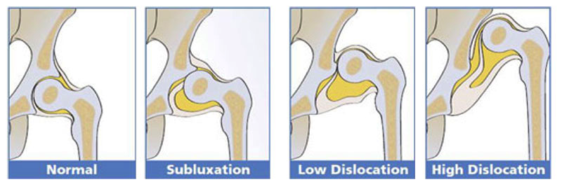
Diagram of Hip Dysplasia Severity
Aside from hip dysplasia, the other indication for this procedure is known as acetabular retroversion. In this uncommon condition, the hip socket may be normally shaped, but is facing the wrong direction (usually too far backwards). This has recently been considered a form of femoro-acetabular impingement (FAI), where in some individuals, the parts of the femur and the acetabulum that normally move smoothly without colliding actually bump into each other, and this can result in restriction of movement and may cause damage at the margin of the hip socket to a structure called the acetabular labrum. It is thought that damage to the labrum by this mechanism is one possible cause for a hip in a young patient may become arthritic in middle-age.
The information above is general. All surgical procedures involve some risk. If you would like advice on your specific condition, please contact the office of Mr Daniel Robin, Melbourne Orthopaedic Surgeon.
03 9044 4555